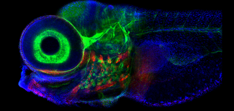
Zebrafish are a scientific wonderfish. They have Wolverine-like regeneration abilities–and can almost entirely regrow their spinal cords after damage. They also give scientists insight into some of the animal brain’s most primal states. While working with week-old zebrafish larvae, a team of scientists decoded how the connections made by a network of neurons in the brainstem guide where the fish looks. They also created a simplified artificial circuit that can predict visual movement and activity in the animal’s brain. This discovery sheds light on how the brain handles short-term memory and could lead to some new ways to treat eye movement disorders in humans. The findings are detailed in a study published November 22 in the journal Nature Neuroscience. It also comes with a striking image taken with a microscope, with vibrant colors that show off the brain regions that are controlling eye movements.
Shifty eyes and changing brain states
Animal brains are constantly taking in a wide variety of sensory information about the environment, even when we don’t consciously realize it. This data is often changing from one moment to the next and the brain faces the challenge of retaining these quick, little kernels of information for long enough to make sense out of them. For example, it must link together what a set of mysterious sounds might be or allow an animal to keep its eyes directed to an area of interest like prey or a potential threat lurking in the distance.
“Trying to understand how these short-term memory behaviors are generated at the level of neural mechanism is the core goal of the project,” study co-author and Weill Cornell Medicine physiologist Emre Aksay said in a statement.
[Related: How animals see the world, according to a new camera system.]
To decode the behavior going on in these dynamic brain circuits, neuroscientists build mathematical models that describe how the state of a system changes over time and where that current state determines the circuit’s future states according to a set of rules. One of the brain’s short-term memory circuits will remain in a single preferred state, only until a new stimulus comes along. When that new stimulus appears, the circuit will settle into a new activity state. In the visual-motor system, each one of these states can store the memory of exactly where an animal should be looking.
However, questions remain about the rules and parameters that help set up that type of shifting system. One possibility comes down to the anatomy of the circuit–the connections that form in between each neuron and how many connections they make up. A second possibility is the physiological strength of those connections. This strength is established by several factors, including the amount of a neurotransmitter that is released, the type of receptors that catch the neurotransmitters, and concentration of those receptors.
Building a neural circuit from scratch
In this new study, the team sought to understand what contributions the circuit anatomy made in the visual system. When they are only five-days-old, zebra “fishlets” are already swimming around and hunting prey. Looking for something to eat involves sustained visual attention and the brain region that controls eye movement is structurally similar in both fish and mammals. However, the zebrafish system contains only 500 neurons. By comparison, the human brain has roughly 100 billion neurons.
“So, we can analyze the entire circuit—microscopically and functionally,” said Aksay. “That’s very difficult to do in other vertebrates.”
[Related: Why do we send so many fish to space?]
While using several advanced imaging techniques, the team identified the neurons that participate in controlling the zebrafish’s gaze and how all of these neurons are wired together. They found that the system consists of two prominent feedback loops. Each of these feedback loops contains three clusters of tightly connected cells. Using this set up, they built out a computer model of what is going on in this part of the zebrafish’s brain.
When the team compared the artificial network that they built with physiological data from a real zebrafish, they found that their fake network could accurately predict the activity patterns.
“I consider myself a physiologist, first and foremost,” said Aksay. “So, I was surprised how much of the behavior of the circuit we could predict from the anatomical architecture alone.”
[Related: Scientists mapped every neuron of an adult animal’s brain for the first time.]
Future applications
In future studies, the team plans to explore how the cells in each cluster contribute to the circuit’s behavior and whether the neurons in the different clusters have specific genetic signatures. This kind of data could help clinicians to therapeutically target the cells that might be malfunctioning in human eye movement disorders. Strabismus occurs when both eyes don’t line up in the same direction and results in “crossed eyes” or “walleye.” The disorder nystagmus presents as fast, uncontrollable eye movements, sometimes called “dancing eyes.”
The findings also provide scientists with a way for unraveling the more complex computational systems in the brain that rely on short-term memory, like those that understand speech or decipher images.


